Joint and Body Pain Treatment in Jersey City
Arthritis
Background: Rheumatoid arthritis (RA) is a chronic systemic inflammatory disease of undetermined etiology involving primarily the synovial membranes and articular structures of multiple joints.
Frequency: In the U.S.: Occurrence rate is approximately 1% in the U.S. The range is from 0.5% to over 5% with some ethnic variation.
Internationally: Similar to the U.S.
Mortality/Morbidity: Mortality from RA is primarily related to the patient's overall deterioration in health, well being and functionality. RA patients become susceptible to infection and secondary organ dysfunction, including lung disease, kidney disease and GI hemorrhage.
Sex: Female to male ratio is approximately 3:1.
Age: Age of onset is usually between 25-50 years. The disease can occur at any age but tends to peak in the fourth and fifth decades of life. The pediatric form of RA is Juvenile Rheumatoid Arthritis (JRA), which is characterized by onset prior to 16 years of age and includes 3 categories of disease: polyarticular (multiple joints), pauciarticular (less than four joints) and systemic (i.e., high fever, rash and organ involvement).
Clinical
History: RA is usually a disease of insidious onset, although it can be abrupt. The diagnosis is typically made when four out of seven qualifying criteria established by the American Rheumatism Association are met. These qualifying criteria are as follows:
- Morning stiffness lasting greater than one hour before improvement
- Arthritis involving three or more joints
- Arthritis of the hand, particularly involvement of the proximal interphalangeal joint (PIP), the metacarpolphalyngeal joints (MCP) or wrist joints
- Symmetric involvement of the same joint areas bilaterally (i.e., both wrists, symmetric PIPs and MP joints)
- Positive serum rheumatoid factor
- Rheumatoid nodules
- Radiographic evidence of rheumatoid arthritis
- Other contributing history includes the following:
- General malaise
- Weakness
- Fever of undetermined etiology
- Weight loss
- Myalgias
- Tendonitis
- Bursitis
- Physical:
- Joint involvement is typically polyarticular and symmetrical, usually sparing the distal interphalangeal (DIP)joints. Joint involvement and inflammation is evidenced by the following:
- Edema
- Effusion
- Warmth
- Tenderness to palpation
- Destruction (a late finding)
- Subcutaneous rheumatoid nodules, "swan neck" deformities, "Boutonniere" deformities, ulnar deviation of fingers at MCP joints (late findings)
- Bursitis
- RA is a diffuse systemic disease involving many areas of the body. The presenting complaint may be remote from a joint or may involve inflammatory symptoms at a joint.
Causes:
- The cause of rheumatoid arthritis has not been elucidated.
- Associated factors may include:
- Genetic predisposition
- Female sex
- Psychological stress
- Immune response
- Hormone interaction
- Work-up
Lab Studies:
- Complete Blood Count (CBC) to detect
- Anemia thrombocytosis
- The erythrocyte sedimentation rate (ESR)
- Serum rheumatoid factor (RF)
- Antinuclear antibodies
Imaging Studies:
- X-Rays
- CT Scan
- MRI
- Thermography
- Joint Fluid Analysis
- Follow-up
Deterrence/Prevention: As the cause of RA is unknown, prevention of the disease has not been established.
There are many suggested approaches to prevent or minimize recurrences or flares. These include proper nutrition, relaxation, low-impact exercise, flexibility exercises, yoga, tai chi, counseling, meditation, hydrotherapy and stress-reduction.
Prognosis: Prognosis in RA is extremely variable.
There is some evidence to suggest that the onset of disease (rapid vs. insidious) may predict the progression of disease. Patients with rapid onset of disease may show better remission than those with insidious onset. Prognosis is worse with large joint involvement.
Patient Education:
Patients with RA require a great deal of education regarding the need for lifestyle changes to prevent exacerbations, to preserve mobility and functionality, and for appropriate pain management.
Specific education for the ED visit is focused on the exacerbation or focal problem that the patient presents with.
Head
Headache Definition: A pain in the head from any cause. See also benign headache; migraine headache, classical; migraine headache, common; tension headache; and the cluster headaches documents.
Alternative names: Cephalalgia; pain in the head.
Considerations: Tension headache and migraine headaches account for 90% of all headaches.
Types:
- Tension: muscle contraction
- Vascular: migrain headache or cluster headache
- Combination: tension and vascular
A headache that signals a potentially serious problem is one that:
- Involves sudden, violent pain
- Could signal an aneurysm
- Gets worse over time and includes other symptoms such as nausea and vomiting, speech changes, personality changes, etc.
- Although rare, it could be a brain tumor or a TIA ("mini-stroke")
- Includes nausea, vomiting, fever, and a stiff neck
- It could be a sign of meningitis
Common causes:
Tension headache is a common headache pattern that may or may not be associated with psychosocial stressors.
Migraine headaches, which are often preceded by fatigue, depression, and visual disturbance (light flash, loss of peripheral vision, etc.).
Cluster headaches, which are a variation of the migraine, are characterized by:
- Pain that occurs mostly in men, while typical migraines are more common in women.
- Pain that is often situated behind an eye and usually the same eye.
- Pain that comes on very suddenly and without warning.
- Pain that peaks within 5 to 10 minutes and disappears in less than an hour.
- Pain that is often triggered by alcohol.
- Pain that will awaken a person from sleep and occur several times a day for weeks and then stop.
Other causes of headaches:
- Sinusitis
- Fever
- Alcoholic hangover
- PMS
- Anxiety
- Dental problems like toothaches
- Tumours
- Trauma
Diagnostic tests that may be performed include:
- Head CT scan
- Head MRI
- Sinuses X-rays
- Temporal artery biopsy
- Lumbar puncture
Treatment:
- Medication
- Physical medicine rehabilitation
- Psychotherapy
Head Injuries
Definition: Trauma to the head.
Alternative names: Brain injury; concussion - first aid; injury to the head.
Considerations: The signs and symptoms of a head injury may occur immediately or develop slowly over several hours.
Most head injuries are minor. The skull provides the brain with considerable protection form injury. Most head injuries are mild, but head injury may be a serious problem when it occurs. Accidents are the leading cause of death or disability of men under age 35, and over 70% of accidents involve head injuries and/or spinal cord injuries. Common causes of head injury include traffic accidents, industrial/occupational accidents, falls, physical assault, and accidents in the home.
If a child begins to play or run immediately after getting a bump on the head, serious injury is unlikely. However, the child should still be closely watched for the next day, since sometimes symptoms of a head injury are delayed.
When encountering a victim of a head injury, try to find out what happened. If the victim cannot tell you, look for clues and ask witnesses.
Even if the skull is not fractured, the brain can bang against the inside of the skull and be damaged. If there is bleeding inside the skull, complications may follow.
Injury or trauma to the head can result in:
- Concussion--the head sustains a hard blow
- Intracranial hematoma--blood vessel ruptures between the skull and the brain (see subdural hematoma)
- Skull fracture
Symptoms:
- Altered level of consciousness
- Bleeding
- Breathing slowed
- Confusion
- Convulsions
- Fracture in the skull
- Fluid drainage from nose, mouth, or ears (may be clear or bloody)
- Headache (may be severe)
- Increased drowsiness
- Initial improvement followed by worsening symptoms
- Irritability
- Loss of consciousness
- Personality changes
- Restlessness
- Slurred speech
- Stiff neck
- Swelling at the site of the injury
- Vision difficulties
- Vomiting (may be severe and persistent)
- Wound in the scalp
- Pupil changes
First aid:
Treatment varies according to the severity of the injury, type and location of injury, and development of secondary complications. For mild head injury, no specific treatment may be needed other than observation for complications. Over-the-counter analgesics may be used for headache. Aspirin is usually discouraged because prolonged use increases the risk of bleeding.
For moderate to severe head injury, urgent treatment is required. The following first aid treatment is indicated if the victim is comatose or symptoms are severe.
- Check the victim's airway, breathing, and circulation. If necessary, begin rescue breathing and CPR.
- If the victim's breathing and heart rate are satisfactory but he or she is unconscious, treat him or her as if there is a spinal injury. Stabilize the head and neck by placing your hands on both sides of the victim's head, keeping the head in line with the spine and preventing movement. Wait for medical help.
- Unless there has been a skull fracture, attempt to stop any bleeding by firmly pressing a clean cloth on the wound. If the injury is serious, be careful not to move the victim's head. If blood soaks through the cloth, don't remove it; just place another cloth over the first one.
- If you suspect a skull fracture, do not apply direct pressure to the bleeding site and do not remove any debris from the wound. Cover the wound with sterile gauze dressing and get medical help immediately.
- If the head wound is superficial, wash it with soap and warm water and pat dry.
- If a victim is vomiting and you don't suspect a spinal injury, turn his or her head to the side to prevent choking. Children often vomit once after a head injury. But even if the child does not vomit again and is not behaving differently, contact a doctor.
- Apply ice packs to swollen areas.
- Over-the-counter pain medicine usualy helps reduce headache.
- Over the next 24 hours, observe the victim for any signs of a serious head injury. During the night, awaken the victim every 2 to 3 hours and check for alertness. Ask the victim specific questions, such as an address. If the victim becomes unusually drowsy, develops a severe headache or stiff neck, vomits more than once, or behaves abnormally, get medical help immediately.
- Refrain from vigorous activity for 24 hours after a serious head injury.
Do not:
- DO NOT remove the helmet of a victim if you suspect a serious head injury.
- DO NOT wash a head wound that is deep or bleeding profusely.
- DO NOT remove any object sticking out of a wound.
- DO NOT move the victim unless absolutely necessary.
- DO NOT shake the victim if he or she seems dazed.
- DO NOT let other, more obvious, injuries distract you from the head injury.
- DO NOT pick up a fallen child with any sign of head injury.
- DO NOT consume alcohol within 48 hours of a serious head injury.
Call immediately for emergency medical assistance if:
- There is severe head or facial bleeding.
- There is a change in the victim's level of consciousness (such as confusion or lethargy).
- There is any cessation of breathing.
- You suspect a serious head or neck injury.
Prevention:
- Always wear a helmet when biking.
- Make sure that children have a safe area in which to play.
- Provide adequate supervision for children of any age.
- Be visible. Don't ride a bike at night.
- Obey traffic signals when riding a bike. Be predictable so that other drivers will be better able to determine your course.
- Use appropriate safety equipment (such as hard hats, bicycle or motorcycle helmets, and seat belts) when involved in activities that could result in head injury.
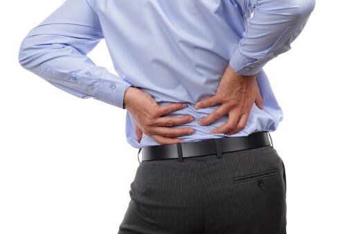
Trunk - Lower Back Pain
Of the numerous musculoskeletal disabling conditions, the complaint of low back pain is undoubtedly predominant. Estimates of hours lost to industry, hours of disability, and money paid for medical care and disability compensation is astronomic. There are as many concepts of the mechanisms and causes of low back pain as there are advocates of numerous forms of treatment. Currently there is no one accepted mechanism that has full acceptance of low back pain.
Functional Unit
The vertebral column, which in its lumbar area is the site of pain, can best be evaluated by understanding of the functional units that comprise the vertebral column. The functional unit is composed of two segments: the anterior portion, which functions in directional guidance.
Anterior Portion:
The weight-bearing portion of each functional unit is the anterior portion, which is comprised of two vertebral bodies separated by a hydrodynamic shock absorber, the intervertebral disk.
The intervertebral disk is a fibrocartilaginous body securely connecting two adjacent vertebral bodies. The individual disk comprises three parts:
Figure 19a: Functional vertebral unit. View of the vertebral body, the posterior articulations (facets), the pedicles,
Figure 19b: Lateral view demonstrating the intervertebral disk and its relationship to the components of the unit.
Figure.20: The functional unit of the spine in cross section: A, Lateral view; B. Shows pressure within disk, which forces vertebrae apart, and the balancing force of the long ligaments; C, Gliding motion of the plane of the facets.
Aging plus multiple microtrauma causes the cartilage plates to become thinner and the posterior aspect of the annulus to become fragmented. There is some invasion of granulation tissue from the vertebral body along with gradual loss of water and the nucleus loses its ability to bind water; thus intradiskal pressure decreases. Further aging or trauma causes a decrease of protein polysaccharide concentration and the disk becomes more fibrous, inelastic, and inert hydrodynamically.
Figure.21a: Annulus fibrous. Above, Layer concept of annulus fibrous.
Figure.21b: Circumferential annular fibres about the centrally located pulpy nucleus (nucleus pulposus).
Degenerative changes decrease the efficiency of the intervertebral disk (Fig.22). These changes may be due to aging, a genetic precursor, or chemical or microtraumatic factors. The hydrodynamics become impaired.
Figure.22: Disk degeneration. Left, Normal disk with intact nucleus and annular fibres. The space is normal(N). Right, Degenerated disk with the nucleus outside its boundary and fragmented annular fibres and narrowed space (D).
The disk functions as a hydraulic shock absorber. The pressure within the nucleus pushes the vertebrae apart and annual fibres pull them together (Fig.23). As weight is added to the unit, the vertebral bodies approximate by deforming the nucleus. Upon release of the compressing force, the nucleus regains its resting form. Flexion, extension, and some rotation is permitted by this nucleus deformation. Compression tests have confirmed that forces will fracture the vertebrae before damaging the disk.
Figure.23: Left, Normal nonweightbearing disk; Center, Deformation of nucleus reacting to compression; Right, Deformation of nucleus permitting flexion or extension.The posterior longitudinal ligament reinforces the posterior aspect of the intervertebral disk. In the lumbar region the ligament tapers to become partial and, thus, affords inadequate protection to the contents of the spinal canal and the intervertebral foramina (Fig.24).
Figure.24: Inadequacy of the posterior longitudinal ligament in the lower lumbar segment, therefore decreasing the protective effect in the L4, L5, and S1 region. Tight, Disk herniation may bulge into the spinal canal, a.
Flexion, extension, and rotation is permitted because of the obliquity and the elasticity of the annular fibres. Rotation of the vertebral column is restricted by the limit of extensibility of the annular fibers.
Extensive rotation exceeding this limit may be a contributing force to disk damage, herniation, and the deterioration.
Nutrition of the intervertebral disk remains an enigma. In the neonatal period many small blood vessels penetrate the vertebral end plates and supply the disk. These blood vessels are obliterated in adolescence and the disk becomes avascular. Nutrition of the disk by osmosis has been disproved and the current accepted method of nutrition is by imbibition. This forms a colliod imbibition pump. As the disk imbibes, its size increases and causes the annular fibres to become taut. Equilibrium is reached by the hydraulic pressure exerted by the vertebral end plates above and below and by the encircling annular fibers.
Diffusion of solutes occurs via the central portion of the end plates and through the annulus (Fig.27). Increased intradiskal pressure probably also forces fluid through minute formina in the end plates. When pressure is released or decreased, fluid returns into the disk by imbibition.
There are numerous marrow spaces immediately under the endochondral plate that connects the bone blood supply to the disk and allows diffusion of the solutes. These spaces are more numerous in the annular region than in the nucleus and more numerous in the posterior position of the disk. This factor possible explains early degeneration of the nucleus and of the posterior aspect of the disk. Solutes containing glucose and oxygen enter by the way of the end plate and sulfates that from glucosaminoglycans enter by the way of the annulus (Fig.27).
Currently the disk is not considered to have innervation within its substance. Numerous investigators have traced nerves of various stages of myelinization and demyelinization to the periphery of the annulus, but no nerves have been verified to enter within the substance of an intact normal disk.
Figure.27: Disk nutrition through dissusion. Diffusion of solutes occurs through the central portion of the end plates and through the annulus. Marrow spaces exist between circulation and hyaline cartilage and are more numerous in the annulus than in the nucleus. Glucose and oxygen enter via the end plates. Sulfate to form glucosaminoglycans enters through the annulus. There is less diffusion into the posterior annulus. (B.V. - blood vessels)
These facets are composed of articular cartilages on their opposing surface and have a capsule, synovium, and synovial fluid. By their shapes and verticality they articulate on each other to permit specific directions of motion and to deny or modify opposing directions of motion. Becuase of their sagittal vertical plane in the lumbar spine, the facets permit flexion and extension but deny or restrict lateral flexion and rotation (Fig.29).
Compared to the anterior joint of the intervertebral disk space, they are not weight bearing. It has been considered that they can support approximately 10 to 12 percent of body weight in the extended spine decreasing to no weight bearing with any significant degree of forward vertebral flexion. In lumbar hyperextension, laterla flexion and rotation can be completely prevented in this posture. The vertebral facets are in complete opposition in a "locked" position. With any degree of forward flexion, separation of the facets occur, thus permitting some degree of laterla flexion and some degree of rotation.
Figure.29: Facets in the lumbar spine. A, Separation of the facets in forward flexion; B, Opposition of the facets in the physiologic lordotic posture; C, Approximation and opposition of the facets on extension and hyperextension.
The posterior wall of spinal canal, the anterior wall of the pedicles, and the lamina are covered by the yellow ligament (ligamentum flavum).
The yellow ligament is an elastic logitudinal ligament extending the length of the vertebral column comprised exclusively of yellow elastic fibers. Its function has been considered to be prevention of the redundant capsule from becoming impinged between adjacent joint surfaces during extension of the spine and limitation of capsular bulging into the spinal canal during other movements of the vertebral column.
Completeting the posterior arc of the vertebral body are the transverse processes and the posterior superior spine upon which attach supporting intervertebral ligaments and the intervertebral muscles that activate vertebral column motion.
The intervertebral foramina are formed by two superior and inferior adjacent pedicles. Anteriorly are the vertebrae bodies, the intervertebral disks, and the posterior longitudinal ligament. Posteriorly are situated the facets, their capsules, and the yellow ligament. Through these foramina emerge the nerve roots, their dural sleeve, and the recurrent nerve of Luschka. The nerve roots descend at the cauda equina.
Erect Balance and Stance
Erect balance requires biomechanic balance. Since the vertebral column is flexible, it would appear that muscular effort is necessary to maintain column relationship to the center of gravity. If this were true, muscular fatigue and collapse of the erect column would ensue. Nature has prevented this total dependence on muscular balance by permitting the column to be supported on ligamentous structure. The lumbar curve extends into greater lordosis, placing its support upon the anterior longitudinal ligament and posteriorly upon the facet joints
The hip joints can be immobilized in the extended position by virtue of the ligamentous bands within the anterior capsule of the hip joint. With the body weight "leaning" against this iliopectineal band (the Y ligament of Bigelow) the hip can remain extended with no muscular activity. The knee joint is prevented from hyperextending by virtue of the posterior popliteal capsular tissues. The knee can thus be maintained in the extended position by ligamentous support requiring no muscular effort.
Only the ankle cannot be immobilized and supported by ligamentous tissue. The ankle, however, can be maintained in the erect weight bearing position with alternating isometric contraction of the anterior dorsiflexors and the posterior gastrocnemius-soleus muscle groups.
Since forward flexion of the lumbar spine is essentially only a reversal of the lumbar lordosis into a slight lumbar kyphosis, full body flexion requires simultaneous rotation about the hip joints of the pelvis as the lumbar spine flexes. The synchronous lumbar spine flexion with pelvic rotation is aptly termed lumbar-pelvic rhythm. For every degree of lumbar spine flexion there is a proportional rotation of the pelvis.
Because the erect stance is essentially a posture dependent upon ligamentous support, muscular support must be elicited the instant the body flexes forward anterior to the center of gravity. As the body flexes forward, the erector spinae muscles elongate to decelerate forward flexion. Insofar as muscular elongation which performs deceleration is a learned reflex, its smoothness and efficiency requires neuromuscular coordination. Poor training or distraction during this motion will interrupt the smooth efficiency onforward flexion. Poor body condition may also, by increasing the fibrous elements of the muscular system, limit the degree of extensibility.
Once the body reaches full forward flexion it is again restricted by ligamentous connective tissue.
To return to the full erect posture the extensor muscles must now shorten and function as acceleration or body elevation until the full erect posture of ligamentous support is achieved.
The return to erect posture must also accomplish the reverse of the flexion lumbar-pelvic rhythm. The lumbar lordosis must be gradually resumed as the pelvis is derotated. Here again, precise coordinated neuromuscular effort must exist and requires training, practice, and no interference by distraction.
Insofar as the posterior articulations, the facets, guide the direction of lumbar flexion and extension and deny lateral flexion and rotation, this direction of reextension must be carefully observed.
In the flexed position, i.e., the body bent forward and the lumbar lordosis reversed, the facets are separated, thus permitting some degree of lateral flexion and rotation. As lordosis is achieved, the facets approximate and, ultimately, as hyperextension is achieved, are in direct contact and prevent any lateral or rotation movement.
In the forward flexed position, the separation of the posterior facet joints place all the limitation of rotation of the functional unit upon the intervertebral disk. It is probable that in faulty rotation with the body flexed forward disk injury occurs.
Therefore, any of the following can result in painful disability: violation of proper lumbar-pelvic rhythm, either in the act of forward flexion or reextension to the erect posture; faulty neuromuscular deceleration or acceleration; or violation of the proper direction of reextension mandated by the plane of the facets. The following factors are related to proper neuromuscular coordination: proper training, constant practice, good conditioning, and no diversion such as can be expected from anxiety, anger, haste, or distraction.
Site of Pain
Since faulty action of the functional vertebral unit can result in pain, it is necessary to elicit the tissues within the unit which, when irritated, can initiate the sensation of pain. Within the functional unit, the following should be noted.
- The anterolongitudinal ligament is sparsely innervated, but chemical, electrical, or mechanical irritation of this ligament does not evoke any significant pain sensation.
- Vertebral bodies are a site of pain as is evidenced in fracture, metabolic disorder, or metastatic disease. The pain from the bodies themselves is dull, vague, usually nonradiating, and not significantly related to motion or position. Nocturnal pain in the reclining position is a symptom that alerts the examiner to suspect disease of malignancy or metabolic origin and initiates appropriate studies.
- The young undamaged intervertebral disk with no evidence of degenerative changes is Vascular, aneural, and, therefore, insensitive. No pain is elicited when performing a diskogram, which requires penetration of the pulpy nucleus with a needle. Injection of a small quantity of material without undue pressure causes no discomfort other than a mild low backache. In jection of an anesthetic agent such as Novocaine does not alter a residual low backache; therefore, intradiskal innervation is verified and altered dynamics must be assumed. Injection of material into an abnormal disk causes severe low back pain with radiation of leg pain.
- The posterolongitudinal ligament has copious innervation of unmyelinated nerves with sympathetic fibers. The nerve supply has been firmly established to be the recurrent meningeal nerve of Luschka. Pain can occur from irritation of these innervated tissues.
- The nerve of Luschka also innervates the aural sheath of the nerve root as it emerges through the intervertebral foremen. The nerve root and its aura are another source of pain within the functional unit. The intrinsic nerve supply from the meningeal ramus has several branches that run through the foremen: one towards the posterior longitudinal ligament, one to the aura mater, and one within the epidural tissue. There are numerous fibers that run longitudinally on the dorsal surface or within the dorsal aura but none are found on the ventral surface or intradurally. The lack of nerve supply to the ventral aspect of the aura possibly explains Falconer and associates] in their claim that the aura was insensitive.
- The yellow ligament is exclusively yellow elastic connective tissue and is devoid of any innervation; therefore it is insensitive.
- The interspinous ligaments are also innervated and can, when inflamed, cause local as well as referred pain. Kellgren injected irritating substance alongside of the spinous process of the first sacral vertebra and caused pain of a sciatica nature radiating down the leg. His injections apparently were into the ligament, partially into the muscle and may have been into the periosteum.
- Stimulation of deep muscles around a spinal joint has been confirmed to cause referred pain. The multifid muscles contract in an attempt to splint the lumbosacral spine in painful low back syndromes. The basis of deep muscle contraction may well result from irritation of the recurrent nerve of Luschka, from the facet joints, from the posterior longitudinal ligament, or even from the protrusion of a damaged disk. If excessive muscular tension can cause symptoms of low back and referred leg pain it can, in part, explain aggravation of symptoms from anxiety, anger, tension, and even chilling as well as faulty use of the back.
- The posterior articulations (facets) are synovial joints and are a site of pain from injury, inflammation, or misuse. The facets are innervated by the articular branch ofthe posterior primary division ofthe nerve root.
In summarization, the tissues capable of causing pain are the posterior longitudinal ligament, the nerve root and its aura, the posterior articulations (facets), the ligaments, and the musculature of the vertebral column.
The examiner should appreciate and determine which of these tissues is the site of pain and the mechanism by which these tissues are being irritated, thus initiating the pain cycle. Assuming that pain mediates through peripheral nerves and ascends the cord, then the mechanism of chemical, electrical, thermal, or mechanical nociceptive stimuli, which is ultimately interpreted as pain from these sites of painful stimuli, form the basis of clinical evaluation of the patient complaining of low back pain.
Low Back Pain
Low back discomfort may be categorized as being either static or kinetic.
In abnormal posture with excessive lordosis there is weight bearing upon the posterior articulation. The foramina are narrowed and the posterior intervertebral disk is compressed and may protrude to encroach upon the posterior longitudinal ligament or laterally into the foremen to compress the aural sac of the nerve root. If any of these tissues have not been previously irritated or inflamed by trauma or stress, no pain may result, but, if the tissues have been sensitized, pain may result.
It has been demonstrated that merely maintaining a forward flexed erect posture of 30 degrees markedly increases intradiskal pressure and demands excessive muscular effort; therefore, this prolonged daily posture may be an incriminating factor in low back pain.
In faulty kinetics with resultant pain several aspects must be considered: (1) the tissues, ligaments or muscles may be restricted in their elasticity, thus limiting full flexibility; (2) the posterior thigh and leg muscles may be restrictive, thus preventing full pelvic rotation and causing the lumbar spine to assume a greater burden; (3) the lumbarpelvic rhythm may be violated, especially in the process of reextension from the fully flexed position.
It may be concluded from the concept expressed that faulty training, poor conditioning, poor musculature, and inflexible connective tissue play a large part in both kinetic and static low back pain. Fatigue, anxiety, anger, and distraction can also play a vital role in impairing proper neuromuscular coordination so vital to proper body mechanics.
It may be concluded from this that faulty training, poor conditioning, poor musculature, inadequate flexibility, fatigue, anxiety, or distraction may result in a painful resultant misuse of the flexion/extension capabilities of the human spine.
Treatment
- Bedrest
- Support - lumbosacra
- Physical Medicine Moodalities
- Rehabilitation Exercises
- Phamocologic Treatment
- Analgesis
- Anti-spasmodic
- Anti-inflammatory
- Epidural Injections
- Surgical Treatment
- Laminectomy Fusion
- Spinal Fusion
- Piriform Syndrome
- Measurement of Pain
Measurement of pain and human reaction to pain remains the greatest challenge to clinical practice. Objectivity, as compared to subjectivity, is uppermost in the mind of the diagnostician and, failing to find an objective basis of pain production, becomes a source of frustration.
Chronic pain localized in the lumbosacral area undoubtedly has an emotional component and all patients presenting with these complaints must have a psychologic evaluation to determine any magnification of the pain claimed by the patient. Intervention, in many instances, must be psychologic as well as an attempt at eradicating or modifying the structural basis of pain. The tests employed by practicing psychologists and by numerous pain clinics have their proponents and are too numerous to list and to evaluate here.
Patient pain drawing is a valuable diagnostic tool. By this technique the area of pain is clarified and is modified by descriptive shading or marking. Such a test facilitates communication otherwise obstructed by language barrier, educational differences, and discrepancy of medical terminology. In patients who are prone to magnify or even falsify their symptoms for whatever gains they see, the organicity or reasonableness of the symptoms is documented.
Pentothal interviews are also valued but used in a different concept of interview. In the somnolent seate, as the patient is lightened from the induced hypnotic state (by way of intravenous peneoehal, movements that induced pain in the wakeful state are performed and the patient's reaction noted. Authenticity of the symptom is given if there is the same response as invoked in the wakeful state, but emotional exaggeration is considered if there is a significant difference. All the precautions in using anesthesia are required so this cannot be performed as an office procedure.
The Minnesota Multiphasic Personality Inventory (M.M.P.I.) is a well-established profile evaluation of a person's personality. Its abuse as well as improper use must be avoided, but complete reliance or infallible interpretation cannot be assumed. The M.M.P.I. categorizes the patient at the time of the test but does not necessarily characterize that patient at the time of initial occurrence of illness or injury.
The Social Readjustment Rating Scale is also a valuable adjunct to evaluation of a patient. The time of disease or injury occurrence has been shown to relate to personal, social, vocational, and economic changes in the patiene's life. High stress versus low stress are thus evaluated and may case an insight into causation of the disability.
Personal interview by a competent and trained psychologist is highly desirable early in the intervention of the illness. When properly invoked, not only accurate diagnosis but also therapeutic intervention results.
The patient with chronic pain is entitled to have thorough medical evaluation to fully evaluate all the organic aspects of the symptoms, and the patient complaining of symptoms of pain muse also be evaluated. Only in all of these aspects can total care of the patient with low back pathology be fully evaluated and treated.
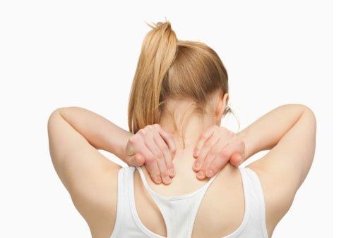
Limbs: Neck and Upper Arm
Pain in the neck, the head, or the interscapular area of the upper extremity (referred there from the neck) is second only to pain in the low back as a medical complaint.
Neck pain may be considered secondary to trauma or it may be appraised as unrelated to trauma. The type of trauma must also be defined. Conditions related to the neck, whether they are post-traumatic or unrelated to trauma, have many similarities but significant differences, and so their evaluation and their management must be clear.


